
Transcatheter Aortic Valve Implantation, commonly known as TAVI, is a minimally invasive procedure used to treat selected patients with aortic valve disease, particularly severe aortic stenosis. Before the procedure, careful planning is essential because every patient’s heart and blood-vessel anatomy is different.
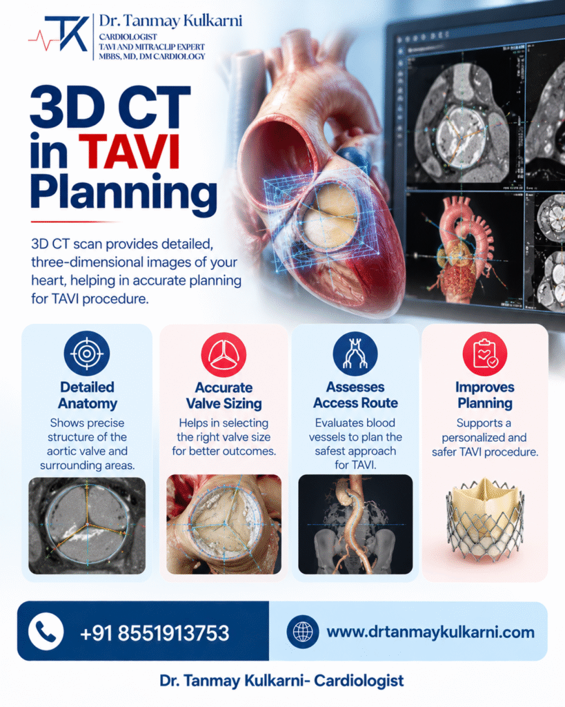
Modern cardiac imaging has made this planning more detailed and individualized. 3D CT in TAVI Planning can provide a comprehensive view of the aortic valve, aortic root, surrounding structures, and potential access vessels. This information can help the cardiac team understand the patient’s anatomy before selecting and performing the procedure.
Dr. Tanmay S. Kulkarni is a cardiologist with expertise in structural heart interventions, including TAVI and advanced CT-based planning.
What Is a 3D CT Scan for TAVI?
A CT scan creates detailed cross-sectional images of the body’s internal structures. For TAVI planning, cardiac CT can provide information about the aortic valve and the anatomy around it.
The images can be reconstructed into different views, allowing the cardiac team to examine structures from multiple angles. This can be particularly useful when the anatomy is complex or when precise measurements are required before a transcatheter valve procedure.
The assessment may include the aortic annulus, aortic root, valve morphology, coronary artery origins, and blood vessels that may be considered for access.
Why Is CT Important Before TAVI?
TAVI involves placing a replacement valve through a catheter rather than removing the existing valve through conventional open-heart surgery. Because the replacement valve needs to be positioned accurately within the existing anatomy, understanding that anatomy beforehand is an important part of procedural planning.
CT imaging can help the cardiac team assess several important features, including:
- Aortic annulus dimensions
- Aortic root anatomy
- Aortic valve morphology
- Valve and surrounding calcification
- Coronary artery height
- Sinus of Valsalva dimensions
- Ascending aorta anatomy
- Potential vascular access routes
These measurements can contribute to decisions regarding the appropriate valve size, procedural approach, and access strategy.
How Does 3D Imaging Help the Heart Team?
Traditional two-dimensional images provide information from individual planes. Three-dimensional reconstruction allows the anatomy to be viewed and analyzed from different perspectives.
This can make it easier to understand relationships between the aortic valve, annulus, coronary arteries, and surrounding structures.
For example, the height of the coronary arteries and the dimensions of the aortic root are important considerations when assessing the potential risk of coronary obstruction. Detailed imaging can therefore contribute to pre-procedure risk assessment.
CT can also help identify the most suitable fluoroscopic viewing angles for the procedure, allowing the heart team to plan the procedure before entering the catheterization laboratory.
Can CT Help With TAVI Valve Sizing?
Yes. CT-based measurements are an important part of TAVI planning and valve selection.
The aortic annulus is not a simple circular structure in every patient. Its shape and dimensions can vary. Detailed CT reconstruction allows measurements of parameters such as annular area and perimeter, which can assist in selecting an appropriately sized transcatheter heart valve.
Valve selection is not based on one measurement alone. The cardiac team considers the complete anatomical picture along with the patient’s clinical condition and the characteristics of the available valve system.
What About Calcification?
Aortic stenosis is frequently associated with calcium deposits around the aortic valve. The location and extent of calcification can be important when planning a transcatheter procedure.
CT can demonstrate the distribution of calcium in the valve and surrounding structures. This information can help the heart team understand anatomical challenges and plan the procedure accordingly.
This can be particularly relevant in patients with heavily calcified valves or other complex anatomical features.
Why Are the Coronary Arteries Assessed?
The coronary arteries originate close to the aortic valve. During TAVI, the replacement valve is positioned within the native valve structure.
Therefore, understanding the relationship between the coronary arteries and the aortic valve is an important component of pre-procedure planning.
CT can help measure coronary artery height and evaluate the surrounding anatomy. These measurements can assist the heart team in identifying patients who may have increased anatomical risk and determining an appropriate procedural strategy.
Does CT Also Assess the Access Route?
Yes. TAVI requires a catheter to travel through a blood vessel to reach the heart. In many cases, the femoral arteries are considered as an access route, depending on the patient’s anatomy and clinical situation.
CT angiography can provide information about the size, calcification, narrowing, and tortuosity of blood vessels along potential access routes.
This helps the heart team assess whether a particular access route is suitable and whether an alternative approach needs to be considered.
What Is AI-Based CT Analysis?
Advanced software can assist with the analysis of cardiac CT images. Depending on the system used, automated or semi-automated tools can help with segmentation, measurements, visualization, and anatomical assessment.
AI-assisted analysis does not replace the cardiologist or imaging specialist. Instead, it can support the clinical team by organizing complex imaging information and facilitating detailed procedural planning.
Dr. Tanmay S. Kulkarni’s website describes experience with AI-driven CT analysis using 3mensio software for TAVI planning.
Is 3D CT Needed for Every TAVI Patient?
The exact imaging protocol depends on the patient’s condition, the suspected valve disease, the planned procedure, and the treating heart team’s assessment.
Not every patient will have the same imaging requirements. CT is generally used as an important component of pre-TAVI anatomical assessment, while echocardiography and other investigations may provide additional information.
The results of all relevant investigations are considered together before a treatment plan is finalized.
3D CT and Personalized TAVI Planning
One of the major advantages of detailed imaging is that it allows treatment planning to be based on the individual’s anatomy rather than relying only on general measurements.
The heart team can assess the valve, annulus, aortic root, coronary arteries, calcification, and vascular access before the procedure. This comprehensive information can help the team prepare for anatomical challenges and select an appropriate procedural strategy.
For patients being evaluated for TAVI, understanding the role of advanced imaging can make the treatment process easier to understand.
TAVI Evaluation With Dr. Tanmay S. Kulkarni
Dr. Tanmay S. Kulkarni is a cardiologist with specialized experience in structural heart interventions, including TAVI. His clinical expertise includes conventional and complex TAVI anatomies, advanced cardiac imaging, and AI-assisted CT analysis.
Patients with aortic valve disease may require a detailed clinical and imaging assessment before treatment decisions are made. The appropriate procedure and imaging strategy depend on individual anatomy, symptoms, overall health, and cardiovascular risk.
A comprehensive evaluation helps the heart team understand the patient’s condition and determine the most appropriate treatment approach.